The Knee Joint
The knee joint is the largest diarthrodial joint in the body. A diarthrodial joint is also known as a "hinge joint." The combined stresses of bearing weight and movement place a lot of strain on the knee. Powerful knee joint extensor and flexor muscles, combined with a strong ligamentous structure, provide a strong functioning joint in most instances. The knee joint is composed of three bones - the femur (or thigh bone), the tibia (or shin bone), and the patella (or kneecap). The parts of the knee joint are subdivided into the tibio-femoral joint which refers to the joint space between the tibia and the femur; and the patello-femoral joint which is the joint space between the patella and the femur. Both of these joints form the knee joint.

Patella
The patella is a sesamoid (floating) bone contained within the quadriceps muscle group and patellar tendon. It's job in the knee joint is to serve as a pulley for the quadriceps muscle group and helps improve the angle of pull. This helps knee extension considerably. It is attached to the tendon of the quadriceps femoris muscle, which contracts to extend/straighten the knee. The vastus intermedialis muscle is attached to the base of patella. The vastus lateralis and vastus medialis are attached to lateral and medial borders of patella respectively.The patella is stabilized by the insertion of vastus medialis and the prominence of the anterior femoral condyles, which prevent lateral dislocation during flexion. The retinacular fibres of the patella also stabilize it during exercise. There are several basic types of abnormalities that may occur with the patella: it may dislocate (slip out of place), sublux (partially slip out of place), fracture, develop degenerative arthritis, or develop a tracking problem.
Dislocated Patella

This type of injury commonly occurs during deceleration. The knee cap slides out of the groove that it is designed for, but does not prohibit motion. Athletes who have structural imbalances and deformities, such as a higher knee cap, have a higher risk of subluxation. If the vastus lateralis is stronger than the inner muscles, the castus medialis, this imbalance may cause pull on the knee cap. Feelings of pressure under the knee cap and pain and swelling behind the knee cap are common symptoms of this problem.

Q Angle

Tibiofemoral Joint |
Patellofemoral Joint
|
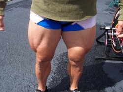
Softball Knee
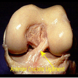 Anterior Cruciate Ligament
|
 ACL Injury
|
Quadriceps Muscle Group
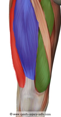
Rectus Femoris
Vastus Intermedius
Vastus Medialis
Vastus Lateralis
Quadriceps Strain